 |
Ultrasound machine with transabdominal and vaginal probes with gel
|
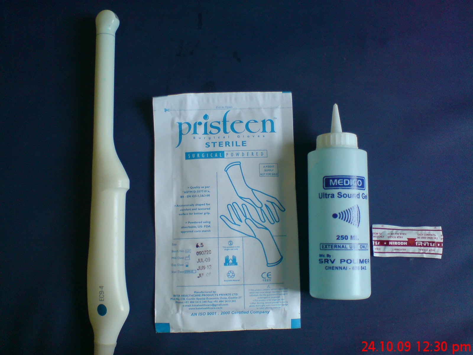 |
| Transvaginal probe with gloves, gel and condom packet |
The development of ultrasonography for study of the fetus and mother is one of the major advances in obstetrics. The first obstetrical application of ultrasound imaging for evaluation of the fetus was by Donald and co-workers in 1958.
The
technique has become a part of obstetrical practice nowadays. With the
aid of a careful sonographic examination vital information about fetal
anatomy, fetal environment, growth and well-being of the fetus can be
noted.
Doppler
methods for measurement of maternal and fetal circulation and three
dimensional views to know maternal and fetal anatomy are also being
used these days.
Safety of ultrasonography:
With the low-intensity sound wave transmission used in real-time imaging, no fetal risks have been demonstrated.
Even
with higher energy intensities used in duplex Doppler imaging also no
embryo or fetal effects are demonstrated. As long as sonographic
contrast agents are not used, there is no hypothetical fetal risk.
The mechanism of action:
In ultrasound the sound waves will play the major role. The
sound waves which will be reflected back from the imaged structure will
form the picture on the screen. The piezoelectric crystals present in
the transducer will convert the electrical energy to high-frequency
sound waves. Gel applied on the skin which is water soluble acts as a
coupling agent.
The sound waves after passing through the tissues will be reflected back to transducer,
converted
into electrical energy and displayed on the screen. Bone like dense
tissue will appear as white structures on the screen such as they
produces high-velocity reflected waves. Amniotic fluid, water etc are
anechoic and they generate only few reflected waves,
so they appear black on the screen.
The picture on the screen appears to move in real-time because the images are generated as quickly as more than 40 frames/sec.
Depending
on the structures to be observed the frequency can be choosed. For
better image resolution high frequency transducers to be choosed. For
better penetration low frequency transducers to be choosed.
To
do abdominal scanning 3- to 5-mHz transducer is normally used but the
obese patients may need a 2-mHz transducer with better penetration to
image the fetus, but the resolution will be decreased. Vaginal
transducers with 7- to 10-
mHz will provide excellent resolution as the structures will be close to the transducer.
The uses of ultrasound:
The
uses of ultrasound in obstetrics can be broadly divided into two, that
is one for accurate estimation of gestational age and two detecting
fetal anomalies.
Estimation
of gestational age by ultrasonography is more accurate than one based
on last menstrual period. When these two are combined even better
results will come. By this either early or too late inductions of labour
can be prevented.
By
doing routine ultrasound examination around 35 to 50 percent of major
fetal anomalies can be identified. With improvement of technology this
percentage is still increasing and can be applied in earlier gestation
age also.
Normally
the anomalies scan is recommended at 18 to 20 weeks as adequate
assessment of fetal anatomy can be best performed after 18 weeks.
Before that time some structures may be difficult to visualize because
of fetal size, position, or movement or maternal abdominal scars or
obesity. The accuracy depends on the skill of the sonographer.
In first trimester:
In the first trimester either transabdominal or transvaginal ultrasonography or both can be used for various indications.
The indications for performing sonography in the first trimester are:
1. For confirmation of intrauterine pregnancy:
if patient having suspicion of having pregnancy, the urine pregnancy
test can detect presence of pregnancy but it will not make out
intrauterine or extrauterine. The transabdominal ultrasound can make out
the gestational sac in the uterus by 6 weeks, and fetal echoes and
cardiac activity by 7 weeks. But with the transvaginal ultrasound, these
can be detected about 1 week earlier.
2. To rule out suspected ectopic pregnancy: by differentiating intrauterine or extrauterine pregnancy, ectopic pregnancy can be ruled out.
3. Identifying the cause of vaginal bleeding:
if a woman presents with abnormal vaginal bleeding the cause can be
identified by the ultrasound examination as it can differentiate between
missed abortion or ectopic pregnancy or other gynaecological causes.
4. To know the cause of the pelvic pain:
by doing ultrasound the causes of pelvic pain can be identified. Which
is either because of impending abortion or due to presence of adhesions
in between pelvic structures etc.
5. To estimate the gestational age:
by doing ultrasound the gestational age can be accurately estimated
especially in the early weeks. By estimating the size of gestational sac
then by calculating the crown-rump length the gestational age can be estimated.
To estimate the crown-rump length the image should be obtained in a
sagittal plane and should not include the yolk sac or a limb bud. If
carefully performed, it has a variation of only 3 to 5 days.
6. To detect the presence of multiple gestations: Multifetal gestation can be
identified in the first trimester by estimating the fetal number, including number of amnions and chorions of multiples when possible, and first trimester is the optimal time to determine chorionicity.
7. For confirmation of cardiac activity:
The transabdominal ultrasound can make out fetal echoes and cardiac
activity by 7 weeks. The transvaginal ultrasound can detect it 1 week
earlier when the embryo is 5 mm in length.
8. In assistance of chorionic villus sampling, embryo transfer:
chorionic villus sampling and amniotic fluid evaluation etc are done to
identify any abnormality in the fetus or in the placenta, these can be
done under direct visualisation with ultrasound guidance more accurately
and with less harm to fetus.
9. To localise or to removal of intrauterine device:
some women may become pregnant with intra uterine device in situ or
some may come with missed threads in these cases ultrasonography is
useful to identify and to remove the device.
10. To evaluate maternal pelvic masses or uterine abnormalities:
ultrasound is useful in detecting the uterine abnormalities or pelvic
masses etc. Actually the first trimester is the best time to evaluate
the uterus, adnexal structures and cul-de-sac etc.
11. In evaluating suspected gestational trophoblastic disease: ultrasonography is useful in detecting the molar pregnancy or partial mole etc. And also useful in follow up of these cases.
12. In detecting genetical abnormalities like Down syndrome:
by doing first-trimester aneuploidy screening by noting the nuchal
translucency along with serum chorionic gonadotropin cases of Down
syndrome can be detected. In detecting trisomy 18 also first trimester
scans are useful.
In second and third trimesters:
The indications of ultrasonography in second and third trimesters are,
1. For estimation of gestational age: ultrasonography is more useful for estimation of gestational age in second and third trimisters.
In
the second and third trimesters the measurements used are the
biparietal diameter, head circumference, abdominal circumference and
femoral length.
Between
14 and 26 weeks, that is in the second trimester the biparietal
diameter (BPD) is usually the most accurate parameter, which will give a
variation of 7 to 10 days. To calculate the BPD distance from the outer
edge of the proximal skull to the inner edge of the distal skull, at
the level of the thalami and cavum septi pellucid to be taken.
Second one is the head circumference (HC) which is more useful than the BPD when the head shape is flattened as in dolichocephaly or rounded as in brachycephaly.
The
femur length (FL) measurement becomes more important as the gestational
age progresses it correlates well with both BPD and gestational age. It
is measured with the beam perpendicular to the long axis of the shaft,
excluding the epiphysis, and it will show a variation of 7 to 11 days
in the second trimester.
Next
is the abdominal circumference (AC), it is the parameter with the
widest variation of 2 to 3 weeks. As it involves soft tissue rather
than bone this much of variation can occur. And also this is the
parameter most affected by the fetal growth. The AC is measured at the
skin line in a transverse view of the fetus at the level of the fetal
stomach and umbilical vein.
As
the pregnancy advances variability of gestational age estimation
increases. By the third trimester, all individual measurements become
less accurate. Estimates are improved by taking an average of the
various parameters the BPD,HC, AC, and FL.
When
combined with estimation from last menstrual period ultrasound
measurements will give more accurate results and also depends on the
efficiency of the sonologist.
For
other fetal structures which will help in detecting abnormalities of
body organs like length of the fetal ears, kidneys, cerebellum, long
bones and feet, and the interocular and binocular distances nomograms
are available.
2. Used in evaluation of fetal growth: by doing ultrasound at regular intervals fetal growth can be evaluated any macrosomia or growth retardation can be identified.
3. In detecting the causes of vaginal bleeding:
in case of vaginal bleeding by doing we can detect cause for it. Either
because of low lying placenta or because of abruption or any other
cause can be identified.
4. With abdominal or pelvic pain:
the causes of abdominal or pelvic pain like abruption placenta or
established preterm labour or rotation of adnexal structures or other
surgical causes can be ruled out.
5. In case of cervical incompetence:
the incompetent cervix can be identified better with trans vaginal
ultrasonography and preventive measures like cerclage can be taken. It
can be useful as adjunct to cervical cerclage also.
6. Determination of fetal presentation:
ultrasound is useful in detecting the presentation of the fetus as
sometimes because of obese abdominal wall or some other reasons
detection of presenting part becomes difficult. If abnormal
presentations are confirmed the plan of delivery can be modified.
7. In case of suspected multiple gestation:
ultrasonography can identify the number of fetuses and the number of
placentas and also the relation of the fetuses which each other so
conjoint twins etc can be ruled out. And the serial growth of the
fetuses can be monitored.
8. As an adjunct to amniocentesis:
the amniocentesis is performed to rule out congenital anomalies and any
metabolic disorders if it is done under ultrasound guidance it can be
done more accurately with less harm to fetal structures.
9. In case of significant discrepancy between uterine size and clinical dates:
some women may calculated their last menstrual period wrongly or
sometimes multiple gestation may be missed during examination, in
conditions like this we can see significant discrepancy between uterine
size and clinical dates, ultrasound will help in correcting the problem.
10. To detect any suspected pelvic mass: ultrasound can detect any associated pelvic mass with pregnancy like uterine fibroid or ovarian tumor etc.
11. Suspected molar pregnancy: ultrasound is useful in detecting the molar pregnancy, the type of molar pregnancy and also in the follow up after treatment.
12. In case of suspected ectopic pregnancy:
normally ectopic pregnancies will not prolong up to second trimesters
but sometimes chronic ectopic pregnancies remain and can cause trouble
later these can be detected by ultrasonography.
13. When fetal death is suspected: in case of any suspicion regarding fetal death it can be ruled out by doing ultrasound and also cause can be found.
14. To rule out uterine abnormality:
uterine abnormalities can lead to abortions or premature labours by
doing ultrasound these can be ruled out and further corrective measures
can be taken.
15. To evaluate fetal well being:
by doing serial ultrasound measurements and by estimating the
biophysical profile by ultrasound we can evaluate the fetal well being.
16. In case of suspected hydromnios or oligohydromnios:
by estimating the amniotic fluid volume, hydromnios or oligohydromnios
can be ruled out. If amniotic fluid index is between 0 to 5 it is taken
as oligohydromnios, in between 6 to 10 it is taken as less liquor, in
between 11 to 15 it is normal liquor, in between 16 to 20 it is excess
liquor, in between 21 to 25 it can be taken as polyhydromnios. If anyone
pocket is more than 8 cm it can be taken as polyhydromnios, if no
pocket is more than 2 cms it can be taken as oligohydromnios.
17. In case of suspected ante partum haemorrhage
: the causes of ante partum haemorrhage like abruption placenta or
placenta previa can be ruled out by ultrasongraphy. In case of suspected
placenta previa in the early gestational age it can be followed to
verify the migration if occurs.
18. As an adjunct to external cephalic version:
when external cephalic version is performed for non cephalic
presentations like breech it can be done under ultrasound guidance.
19. In case of preterm prematurely ruptured membranes or preterm labor: the amount of liquor and the condition of the fetus can be estimated and according to that further plan of action can be made.
20. Abnormal biochemical markers: in case of abnormal biochemical markers, the associated congenital anomalies can be ruled out.
21. To detect fetal anomaly:
the most common malformations can be detected in the second trimester
are cardiac anomalies, doing fetal echo is useful in detecting them. The
second most common are neural tube defects like anencephaly,
menigomyelocele etc can be detected by ultrasound. In case of women
having history of previous baby with congenital anomaly, later
pregnancies should be followed up carefully. Other abnormalities like
diaphragmatic hernia or omphelocele etc can be ruled out.
22. In women with no previous prenatal care:
when a women comes to hospital directly near term or in labor,
ultrasonography will be of great help in knowing the status of the baby
and estimating the gestational age.
23. In induction of labor:
transvaginal ultrasonography is useful in predicting the success of
induction of labor by estimating the cervical changes and calculating
the Bishop score.
No comments:
Post a Comment