Wednesday 8 August 2012

Chocolate cyst or endometrioma


Sometimes when endometriosis affects ovary that can form endometrioma or chocolate cyst,

 
endometriotic cyst usually show some characteristic features like hypo-echoic area with thick wall etc,
and cancerous cysts will have features like thick septa, irregular wall, hetero-echoic areas etc.
usually blood test can detect the levels of CA 125, but it that can be supportive but not confirmatory for diagnosis.
CA 125 levels will be elevated in many conditions other than carcinoma and endometriosis is one of them,
by taking into consideration of ultrasound features and elevated CA 125 levels  possible diagnosis of endometrioma can be made sometimes, but confirmation can be done by histopathological examination only.

as  ovaries are involved in endometriosis by forming chocolate cyst that can also lead to increased frequency of cycles called polymenorrhoea that may also be one cause for early onset of periods.
menorrhagia(excessive bleeding during periods) and dysmenorrhoea(pain during periods) are symptoms of endometriosis,
size of the chocolate cyst of ovary can increase during periods due to accumulation of cyclical blood and after periods there may be some amount of shrinkage due to absorption of serum,


though it is said that some type of foods can affect estrogen levels, theoretically in the pathogenesis of endometriosis these are not stressed.


Normally small cysts can be managed with medicines like analgesics, hormonal therapy like contraceptive pills, progesterone only pills,
as the cyst contains endometrial tissue it can secrete blood periodically, which can cause enlargement of the cyst, so better to take medical treatment like  small dose oral contraceptive pills cyclically or preferably continuously which can cause endometrial tissue atrophy leading to limiting the growth of the cyst.
other medicines like danazol or GnRH analogues etc can also be used.

But for a cyst with size of more than 4 cms  surgical removal is best option, as hormonal treatment in this case is only partially effective and can cause side effects also,
patient can go for laparoscopic surgical approach, as laser surgery and cauterization are less effective and recurrence chance is there , better to go for laparoscopic excision of ovarian adhesions and endometrioma.
Patient can travel but have to be careful and take precautions like, avoid lifting, pushing, pulling weights etc, avoid acts that will increase abdominal pressure like constipation etc, but should be cautious and if pain abdomen etc occurs have to consult doctor immediately.

usually chocolate cyst will rupture because of some strenuous activity or by external pressure on the abdomen etc, by limiting these things we can prevent spontaneous rupture to some extent.


( The viewers are invited to post comments and ask questions related to the topic through comment box.)
 

Emergency contraceptive pill - short term and long term side effects due to repeated usage


The short term side effects of emergency contraceptive pill are,
-they can cause menstrual irregularities like,
if taken in the first half of cycle they can prepone the menstruation, if taken in the second half they can postpone the menstruation.
They can lead to spotting, brownish discharge etc in between periods,
-nausea, vomitings, fluid retention, bloating sensation of the abdomen, breast tenderness, headache, sinus congestion etc symptoms can occur.
-less effective than regular pills with around 13% of failure rate,
-will not protect against sexually transmitted diseases,

Long term side effects:
-Because of repeated usage of pill the pattern of ovulation can change, it may become delayed or gets inhibited which will cause problem in getting pregnant for some months.
-if pregnancy occurs after repeated intake of pill baby may have some kinds of defects,
-repeated usage will decrease the effectiveness of the pill,
-it can affect the growth of endometrium leading to altered menstruation that can lead to amenorrhea or polymenorrhea,
-if used frequently with less than two months interval, that can lead to arrest of growth of ovum leading to formation of cysts that may lead to poly cystic ovarian disease sometimes according to few studies.


( The viewers are invited to post comments and ask questions related to the topic through comment box.)
 

Mittelschmerz

 Mittelschmerz and associated symptoms:
mittelschmerz means ovulatory pain seen on one side of abdomen usually, which can occur due to,
- stretching of capsule of ovary during release of ovum,
-contraction of fallopian tubes to recieve ovum,
-contraction of smooth muscle cells on ovary or of ligaments  due to release of prostaglandins,
-irritation of peritoneum due to blood and fluid released from ovarian follicle,
that pain sometimes can cause nausea,

mild spotting or brownish discharge etc can be seen associated with it due to the decrease in level of estrogen during ovulation,
other symptoms like head ache etc may also be due to release of prostaglandins etc during ovulation,
some women may also experience breast tenderness, bloating sensation, water retention, swelling of genital parts etc,
more intense ovulatory pain can be seen in endometriosis and pelvic inflammatory disease etc conditions.

 if  symptoms subsided soon and if that not severe enough to seek medical advice then no need to bother, if  symptoms persist or become severe better to consult gynecologist once to rule out any other possible cause.

Monday 30 July 2012

Morphea in pregnancy

Morphea  is a localized form of scleroderma which is more common in females than males.
It usually occurs between 3rd to 5th decade, though can occur early also.

There is no much data regarding morphea in pregnancy, according to available studies around 24% of abortion rate reported
And also there is around 71% normal deliveries, so there is no sufficient data to tell exactly the relation between morphea and pregnancy.


as morphea is localized form it affects mainly skin, affecting internal organs is somewhat less, but the risk cannot be completely excluded,

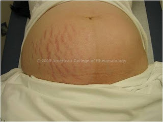
morphea on left side affected the development of  striae of pregnancy and growth to some extent


women with morphea usually have sub-fertility and in case of pregnancy they will be affected more in third trimester usually.
complications that can develop in third trimester:
rapid development of hypertension,
renal failure,
preterm labor,
malabsorption, increased constipation,
growth retardation of the baby, anomalies can also occur because of medicines used by mother etc,
as it affects blood vessels by causing proliferation of connective tissue, inflammatory response and leading to fibrosis of vessels it can affect the growth of the placenta and also its function.

but with proper follow up most of these can be prevented.
 pregnancy can be properly managed with,
-regular antenatal checkups,
-including checking for blood pressure and edema, assessing cardiopulmonary, renal and gastro-intestinal systems, symptomatic treatment for musculoskeletal problems,
-nutritional diet etc.

Saturday 30 June 2012

The causes of false positive pregnancy test


Appearance of positive line in pregnancy test kit in absence of pregnancy is called false positive test result,


That can be seen in conditions like,

-errors of test application,
reading test after suggested 3 to 5 minute window or reaction time,
kit with past expired date

-taking hCG injection as part of infertility treatment,

 
-in some diseases of liver, carcinomas


- medical conditions which will produce elevated hCG leading to false positive test.
choriocarcinoma, 

germ cell tumors,
IgA deficiency,
Heterophile antibodies,
Gestational trophoblastic diseases and neoplasms

Monday 18 June 2012

The possible causes of IVF failure and solutions


The possible causes of IVF(in vitro fertilization) failure or implantation failure are,
1.    In case of PCOD:
 Anovlation is the feature of PCOD. 
And ovulation induction drugs to be used generally.
As there is anovulation, doctors will super ovulate in these patients which will lead to poor quality ovum or
Most of the doctors will think of ovulation hyperstimulation syndrome and they induce less number of ovum. 

Solution:
This can be overcome by careful aspiration of ovum from follicles  and removing follicular cells around it.


2.  Transferring usual number of embryos as in normal patients will not be sufficient in IVF failure cases.

Solution:
So more number of embryos to be transferred in these patients as the chance of high-order multiple pregnancy is less in them.

3.Sometimes patients will have poor ovarian reserve,

Solution:
This can be overcome by aggressive super-ovulation or
by GIFT- gamete intrafallopian transfer.


4. In case of technical difficulties for embryo transfer like cervical stenosis,

Solution: ZIFT- zygote  intrafallopian transfer can be done.

5.Sometimes chromosomal abnormalities will be there in even good looking embryos which lead to failure of IVF.

Solution:
This can be overcome by taking embryo biopsy on 3rd day by laser and transferring the embryo on 5th day.

Monday 28 May 2012

Detection of pregnancy - Urine and blood tests


To detect pregnancy both blood tests and urine tests are available.
 
There are three types of blood tests to detect pregnancy. 
1.       Quantitative blood tests:
pregnancy is generally detected in blood tests by estimating the levels of HCG( Human chorionic gonadotropin).
These can be detected in blood after implantation.
Usually implantation occurs 6 to 12 days after fertilization.
Means as ovulation usually occurs in 12 to 16 days of first day of menstruation, the time of implantation will be around 20 to 26 days from first day of menstruation.
These quantitative tests can detect HCG levels even at 1 mIU/ml.
So by this test we can detect the pregnancy around 25th to 26th day of cycle in case of regular periods.

2.      Qualitative tests:
this tests can detect pregnancy at HCG levels of 25mIU/ml.
 They are less sensitive and by this test pregnancy can be detected after missed period.

3.      Rosette inhibition assay test:
it detects EPF(early pregnancy factor) instead of HCG.
This EPF can be detected in pregnancy within 48 hours of fertilization.
So it can detect pregnancy around 16 to 18 days from first day of periods.
But this test is expensive and available in few centers only.


Urine tests to detect pregnancy:
Urine pregnancy kits usually detect pregnancy at HCG levels of 20mIU/ml to 100mIU/ml.
Their sensitivity depends on the brand and level of HCG they will detect.
They can detect pregnancy at the time of missed period or within 1 week of missed period depending on sensitivity.


Friday 25 May 2012

Itching over abdomen in pregnancy


Itching of the abdomen is a normal finding in pregnancy.
It occurs because of,
-As the uterus grows with the progression of pregnancy, skin stretches to accommodate it  by separation of collagen fibers which will lead to moisture deprivation.
-Changes in levels of hormones during pregnancy is also one cause.


Itching because of stretching is commonly felt on the abdomen, breast, thighs etc. 

But if generalized itching is present it may indicate some underlying problem like cholestatic disease etc.

To get relief from itching,
-          use luke warm water for bath, avoid hot shower baths.
-          mild soap or shower gel to be used, it should be rinsed well and towel off lightly.
-          Then apply cream containing vitamin E and alovera like ‘Elovera cream’.
-          Anti-itch lotion like pink calamine lotion can be used.
-          should not scratch.
-          Choose loose cotton clothes.
-          Avoid going out in hot weather.
-          Excessive weight gain to be avoided.

 
-          Good hydration and balanced nutrition to be maintained.
 -          Avoid continuously sleeping in same position.

Thursday 10 May 2012

Eosinophilia


Eosinophils are the cells produced in bone marrow and found in blood stream and gut lining.

The help in fighting against infections and parasitic infestations.
The normal eosinophil count should be not higher than 350 eosinophils per microL of whole blood.
 If it more than 400 per microL it is called as eosinophilia, means presence of abnormal high amounts of eosinophils in blood and body tissues.


It is seen in conditions like,
Asthma, 
hay fever,
 lung diseases like  Loeffler's syndrome, 
vasculitis as in Churg-Strauss syndrome,   
tumours like lymphoma, 
cirrhosis of the liver, 
in antibody deficiency cases,
 in some skin disorders etc.
If cause is not known it is called as hypereosinophilic syndrome.
The symptoms depends on cause like,
wheezing and breathlessness in asthma,
abdominal pain, diarrhoea, fever, rashes etc in parasitic infestation.
To treat eosinophilia, the cause should be treated,


Wednesday 9 May 2012

Cough and cold in pregnancy


 Though cough and cold are quite common but when they occur in pregnancy they should be treated carefully.
Normal dry cough won’t affect the baby much but if severe cough is there with expectoration it can affect the baby by,
- while coughing increased abdominal pressure will lead to rupture of chorionic membranes and leakage of amniotic fluid sometimes.
- the underlying cause of cough can affect the baby like in case of cough due to some infections, the infecting organism can affect the baby also.
But  there are natural protectors inside the body like, the amniotic fluid will protect the baby from the increased pressure while coughing. And placental barrier to some extent will prevent the entry of organisms from the mother to the baby.
Cough syrups will contain different ingredients meant to reduce particular symptoms of cough like anti histamines, cough suppressants, expectorants, decongestants and analgesics.
 The recommendations during pregnancy are,
-Anti histamines to reduce inflammation, in pregnancy the safe antihistamines are loratadine, doxylamine, diphenhydramine etc.
-Cough suppressants to stop coughing, dextromethorphan is considered to be safe.
-Expectorants to dilute the mucus, guaifenesin is relatively safe in second trimester.
-Decongestants to reduce the congestion of sinuses, pseudoephydrine hydrochloride is considered to be safe.
-Analgesics to relieve the pain, acetaminophen is considered to be safe.
According to above combination the cough syrup can be choosed. But those containing alcohol to be avoided. Along  with this the underlying cause to be treated.
 

The natural remedies for cough and cold are increasing water intake also other fluids like soups, juices, warm tea can be taken which will dilute the mucus secretions.

Inhaling steam also do same thing and also decrease sinus congestion, to treat sore throat you can use lozenges tea with honey also help. By adding moisture to air with humidifier cough can be improved.
If cough is getting increased due to supine position semi-recumbent position can be opted   by putting pillows under head and back.

Monday 30 April 2012

Clinzen in pregnancy


Some people think that clinzen vaginal tablets can lead to miscarriage.
These are frequently prescribed tablets for vaginal infection especially candidiasis.
Clinzen vaginal tablets or cream contain Clindamycin phosphate which is a water soluble ester of the semi-synthetic antibiotic.
It is Category B drug in pregnancy.
There are no adequate and well controlled studies regarding its usage in first trimester of preganacy. It should be used only if clearly indicated.
In second trimester in women treated with clinzen for 7 days abnormal labor reported in 1.1% of women.
As such overall reproductive studies on humans revealed no harm to the fetus. But studies on only one mouse strain reported cleft palate with other strains being not effected.
So, this can be taken as strain specific.
In general hospital practice this is used frequently and not connected to miscarriage.

Saturday 21 April 2012

Vesicovaginal fistula(VVF) - 1


One of the most common causes of urinary incontinence in females is vesicovaginal fistula(VVF). This is most common type of genitourinary fistula.

Definition:


This is a communication between the bladder and the vagina which will lead to escape of urine into the vagina leading to true incontinence.

Etiology:
Both obstetrical and gynecological causes can lead to vesicovaginal fistula.

Obstetrical causes:
Obstetrical causes are responsible for 80 to 90% cases of vesicovaginal fistula in developing countries and 5 to 15% cases in developed countries.

1. Obstructed labor: in case of obstructed labor because of prolonged compression on the bladder base between fetal head and symphysis pubis, ischemic necrosis occurs. Because of this decreased blood supply infection super imposes which will lead to sloughing of tissue resulting in fistula. In this case fistula can develop in 3 to 5 days.

2. Difficult labor: when baby delivery becomes difficult due to big baby or contracted pelvis etc, instrumental delivery may be needed. If forceps are applied especially Kielland forceps, the chances of injury to the bladder is more. 


3. Destructive procedures: in case of difficult labors if baby dies, to take out the baby destructive procedures like craniotomy may be needed. In this case trauma may be caused by instruments used or because of bony spicules of fetal head.


4. Emergency surgeries: in cases of emergency cesarean section for prolonged or obstructed labor, as the bladder will be drawn up in these cases chances of injury is high.
In case of cesarean hysterectomy for rupture uterus the chances of bladder injury is high as here also prolonged labor may led to drawn up bladder.

5. Repeat cesarean section:  because of adhesions caused by previous cesarean section between the bladder and the lower part of uterus, the chances of bladder injury are high.


Monday 16 April 2012

Doppler in Obstetrics and Gynecology


Doppler velocimetry is a noninvasive way of assessing blood flow by characteristic downstream impedance.

Principle :
When sound waves strike a moving target, the frequency of sound waves reflected back is shifted proportionate to the velocity and direction of the moving target. As the magnitude and direction of the frequency shift depend on the relative motion of the moving target, the velocity and direction of the target can be determined.

In obstetrics :
Doppler is used to determine the volume and rate of blood flow through the maternal and fetal vessels.
Here the sound source is the ultrasound transducer, the moving target is the red blood cells flowing through the circulation. The reflected sound waves are observed by the ultrasound transducer.

Uses :
Recommended as possible adjunct to other fetal evaluation techniques.

Antepartum fetal heart rate testing :

 
In detecting the fetal heart rate patterns, accelerations or decelerations in fetal heart rate, used in nonstress test, contraction stress test, biophysical profile etc.

Doppler index:


The umbilical artery systolic/diastolic (S/D) ratio is most commonly used Doppler index.
S/D ratio = maximum(peak) systolic flow/ end diastolic flow
By this the ratio evaluates the down stream impedance of the flow. 
The resistance to umbilical artery blood flow during diastole will be initially high then decreases as the gestational age progresses.
Normal flow :
S/D ratio 4.0 at 20 weeks to 2.0 at 40 weeks.
Abnormal flow:
If elevated 95th percentile of the gestational age
If diastolic blood flow is either absent or reversed.
It indicates extreme downstream resistance, placental dysfunction and fetal compromise with chances of growth restriction.

In estimating fetal maturity:
 
Fetal maturity can be estimated by adding 30 weeks to the date of fetal heart detection by Doppler ultrasound.

In conditions like postterm pregnancy, diabetes, systemic lupus erythematosus, antipospholipid antibody syndrome or in general obstetric population Doppler is ‘not of’ much use.

In Gynecology :
In Gynecology also Doppler is used in different conditions to evaluate the blood flow pattern of different organs and the possible causes for them like,
 
  1. In ovarian and endometrial cancer,
  2.  Pelvic pain,
  3.  Benign and malignant adnexal masses,
  4.  Myometrial invasion of endometrial carcinoma,
  5.  The normal endometrium and benign endometrium disorders,
  6. Normal pelvic blood flow,
  7. Uterine sarcoma,
  8. Uterine myomas.  etc.


Sunday 15 April 2012

Hydrorrhea gravidarum


Definition:
Hydrorrhea gravidarum is defined as “the discharge of a watery fluid from the vagina during pregnancy”. It can also be called as hindwater.
 One more definition is constant or periodic flow of a serous or seropurulent liquid, resembling liquor amnii, from the vagina in pregnancy.

The causes of are:
Rupture of membranes
Hydrosalpinx
Edema of uterine walls
Inflammation of decidual glands
Placenta marginata

Pathology:
Inflammation of deciduae is not uncommon in pregnancy. It occurs usually secondary to slight degrees of endometritis. But  severe degree of endometritis leads to sterility.
The inflamed decidua will be greatly thickened either localized or diffuse and may present with polypoid growths or small cysts.
The rupture of these cystic decidual glands, with sudden gush of fluid from vagina will present as hydrorrhea gravidarum.

Quantity :
Little or more

Time :
Usually occurs after six months of pregnancy

Differential diagnosis:
Liquor amnii
Urine
Local wounds pus discharge
Secretions from local tumors

Dangers :
It can lead to preterm labour

Treatment :
No particular treatment available. Cause to be treated.


Thursday 12 April 2012

RENAL TRANSPLANTATION - PROCEDURE


        Rutherford –Morison incision
        Muscle layers divided
        Inferior epigastric vessels ligated & divided

        Iliac vessels mobilized
        Int .iliac & gluteal branches may need ligation

  Cold kidney is placed in iliac fossa


        Arterial and venous anastomosis  done

        Ureteroneocystostomy  done in extravesical fashion

POSTOPERATIVE CARE 
Foley catheter - Remove on 5th  POD, administer dose of antibiotic

Ureteral stent, if used- Remove 6–12 wk postoperatively in clinic

Drain(s)- Remove when ≤ 30 mL/24 h or in 3 wk if volume > 30 mL/24 h


Tuesday 10 April 2012

RENAL TRANSPLANTATION - RECIPIENT PROCEDURE


Recipient  procedure
  • Endotracheal intubation for GA
  • Establishment of arterial line & CVP monitoring access
  • Pt. is placed in supine position
  • Oblique Rutherford-Morrison incision
  • Ext.oblique, Int.oblique, Transversus abd. muscles divided
  • Inferior epigastric vessels ligated & divided
  • Spermatic cord freed & retracted (round ligament can be divided)
  • Peritoneum pushed medially& upwards
  • Common , Ext.iliac, Int.iliac arteries mobilized
  • Ext. iliac vein mobilized
  • Para psoas gutter developed for final placing of kidney
  • Vascular clamps applied  to Ext/int.iliac artery & Ext.iliac  vein 
  •     Venotomy & arterotomy are given


Back table work for vascular variations

Vascular anastomosis
Renal artery to Ext.iliac artery
                   End to side  fashion with 5-0 or 6-0 prolene
                   ( in End to End fashion if Int.iliac artery is selected )
Renal vein to Ext.iliac vein
                   side to side  fashion  with 5-0 or 6-0 prolene
Kinks and twists are avoided during vascular anastomosis

Reconstruction of urinary tract
  • Orientation of renal vein, artery & pelvis is reversed antero-posteriorly  when opposite iliac fossa is selected

  • Uretero neocystostomy is done in Lich-Gregoir’s  extra vesical  technique  with 3-0 or 4-0 synthetic absorbable  suture material like polyglactide or polydioxanone

  • Placement of Double J stent is optional

  • Wound is closed in layers after fashioning  a drain

Patient in supine position
Foley’ catheter placed

Sunday 8 April 2012

RENAL TRANSPLANTATION - DONAR SCRENING AND OPEN NEPHRECTOMY


Routine live kidney donor screening

 Urinalysis –
  •  Dipstick for protein, blood & glucose
  • Microscopy & culture
  • Protein excretion rate

Assessment of renal function-
  • Estimation /measurement of GFR
Blood tests
Haemogram,coagulation profile
Haemoglobinopathies& G6PD  deficiency (when indicated)

Biochemical profile
       Blood urea, S.Creatinine, S.electrolytes, LFT, FBS, GTT(if indicated)
       Lipid profile,Thyroid profile, Pregnancy test & S.PSA

Virology& infection screen
        Hep.B, Hep.C, Toxoplasma, HIV, HTLV,CMV, EBV 

Cardio-respiratory system
       Chest X-ray, ECG, Stress test, 2D-ECHO(when indicated)

Assessment of renal anatomy& function
       KUB-IVP series, DTPA-Renogram, Renal Angiogram


  • PAC (Pre Anesthetic Checkup) and
  • Pre –Operative  Preparation
                                 ---For both donor & recipient
      
Donor Nephrectomy
  • Open              [ODN]
  • Laparoscopic   [LDN]

Open Donor Nephrectomy(ODN)
  • Endotracheal  intubation for GA
  • Kidney/flank position
  • 11th or 12th  rib incision (with rib excision when needed)
  • Ext.oblique, Int.oblique,Transversus abdominis with fascia divided
  • Retroperitoneum entered
  • Kidney mobilized all around
  • Tributaries of renal vein ligated
  • Renal vein & Renal artery are mobilized up to their origins
  • Ureter mobilized up to pelvic brim and transected
  • Brisk diuresis ensured from the cut ureter
  • Renal artery & Vein are clamped, cut and ligated
  • Kidney is handed over to perfusion team for placing in ice slush & perfusion  with chilled RL & heparin
  • Wound closed in layers with or without a drain


Friday 6 April 2012

RENAL TRANSPLANTATION - HISTORY, SELECTION OF RECIEPIENT


This is guest article by Dr.N.Anil kumar, Urologist.
History and Major landmarks
1933    - First Human Renal Allograft by Voronoy in Ukraine.
1954    - First long term Success with Human Renal   Allograft achieved in Boston.
1958    - First Histocompatibility antigen was  described.
1959    - Radiation was tried for immunosuppression.
1962    - Glucocorticoids became a part of   immunosuppression.
1962    - First use of tissue matching for selection of Donor-recipient pair.
1966  - Direct cross match between Donor  lymphocytes and  recipient serum introduced.
1970’s- Brain Death laws were passed
1978  - first clinical trials of cyclosporine were reported
1981  - Successful use of monoclonal antibodies for renal allograft rejection
1989  - Recombinant Erythropoietin  became available
1995  - Laparoscopic donor nephrectomy was introduced


Urologist’s role -recipient
Ø      Preliminary screening
Ø       Kidney disease recurrence
Ø       Infections
Ø       Active malignancy
Ø       Probable risk factors for perioperative morbidity
Ø       Non-compliance
Ø       Unsuitable conditions for technical success

1. Preliminary  screening :
  • Drug / substance abuse
  • Morbid  obesity
  • Compliance issues
  • Heart disease

2. Kidney disease recurrence
High Risk of Graft Failure
  • FSGS
  • HUS
  • Oxalosis

Potentially treatable
  • Renal Amyloidosis
  • Cystinosis
  • Fabry’s Disease

Graft failure  is rare
  • DM
  • IgA nephropathy

Do not recur in transplanted kidney
  • ADPKD
  • Renal  dysplasia
  • Alport’s without anti BM antibodies

3. Infections
  • Dental sepsis
  • Infection of dialysis access sites
  • Pulm.infections & Tuberculin test
  • Symptomatic cholelithiasis & cholecystitis
  • Recurrent diverticulitis
  • Diabetic foot ulcers
  • UTI
  • CMV, HSV, EBV,  Hep.B, Hep.C, HIV

4. Active  malignancy
  • Invasive cancers- 2-5 yrs of waiting time after last treatment recommended
  • Low grade / Non-invasive cancers-  shorter intervals may be accepted
  • GB-Polyps of >1cm in diameter- cholecystectomy is recommended

5.Probable risk factors for perioperative morbidity & mortality
  • Cardiac risk factors
  • Peptic ulcer disease
  • Significant pulmonary  disease
  • Smoking

6. Unsuitable conditions for technical success
  • Signs& Symptoms of  lower extremity arterial disease
  • H/O abdominal or pelvic vascular surgeries
  • H/O previous vascular thrombosis, antiphospholipid antibodies,  previous transplant renal vein thrombosis
  • Diseases & operations on urinary tract